
With 3D medical imaging advancing, radiologists have come a long way from the early days of using computed tomography (CT scanners) and mammography devices, with new technology enabling them to access new angles and resolutions for improved patient healthcare.
AI is also proving its worth as it automates routine procedures, providing faster image processing for diagnosis and allowing healthcare staff to prioritise and respond to the most demanding and critically-ill patients more efficiently.
However, medical imaging is one of the most common and overused diagnostic techniques used today, and by far the costliest.
The devices are hard to transport, and require a reliable source of power, which limits the use of the technology across rural communities and under-resourced villages in the developing world.
Here we talk to Dr Matthew Morgan, a board-certified radiologist specialising in breast imaging and physician specialist of workflow and decision support at Elsevier, a global information analytics business specialising in science and health.
He acknowledges the concerns regarding healthcare imaging equipment being too expensive as well as trained professionals available to operate the technology.
He said: “Obtaining and operating medical imaging devices is expensive, which inhibits their use in poor areas and developing countries.
“Moreover, experts trained to interpret the imaging [radiologists] are also limited. The combination of these two factors has restricted the benefit of advanced medical imaging to wealthier peoples and countries.”

How will new technology using medical imaging make healthcare more accessible to people across the world?
To make medical imaging more accessible, healthtech companies are designing products by leveraging smartphones to create affordable and portable mobile ultrasound machines.
An example is the Butterfly iQ, which claims to be the first ever handheld whole-body ultrasound system, created by Butterfly Network, a company based in Connecticut.
A clinician can now perform a variety of scans using this one portable device, and since it can be taken just about anywhere, this may be one of the most versatile ultrasounds on the market.
It’s a technique Dr Morgan has also observed, as he said: “One imaging technology that is becoming more affordable is handheld portable ultrasound.
“Smartphone apps and handheld portable transducers allow bedside ultrasound to be nearly as convenient and available as the stethoscope.
“This tool can be used to augment the physician’s physical exam, which is a significant benefit in situations where time is limited and/or where other more advanced imaging equipment is not available.”
Another solution Dr Morgan suggests is utilising AI for earlier and more precise detection, as the opportunities for its implementation in medical imaging are profound.
He said: “Another hope is that AI can act as an adjunct detection tool for alerting and prioritising exams with acute findings.
“While this approach has benefits for technologically advanced countries, it also has the potential to extend the benefits of medical imaging interpretation to under-served populations, where access to trained radiologists may be limited.”
How can medical imaging reach its ultimate potential?
Radiology has become a distinct medical specialty and is now the key diagnostic tool for many diseases, with an important role to play in monitoring treatment and predicting outcomes.
Dr Morgan states: “Medical imaging plays a critical role in nearly all medical diagnosis. It allows physicians to ‘peer inside’ the human body to see what is actually happening.
“Each modality including radiography (x-ray), ultrasound, CT, magnetic resonance imaging (MRI), and nuclear medicine play a unique but pivotal role in identifying and characterising disease.”
Dr Morgan illustrated the wide array of examples that use imaging, from brain imaging that can guide treatment for cancer and seizures, to body imaging, which guides the treatment of infection and trauma.
He explained: “Brain imaging that guides the treatment from cancer to seizures, and functional brain imaging is augmenting our understanding of mental health.
“Imaging of the heart enables the treatment of heart attacks and failing heart valves.
“Body imaging guides the treatment of infection, trauma, cancer, and a host of other conditions.
“Imaging touches nearly every patient in the hospital.”
Radiologists have also had an impact on the advancements of medical imaging methods to include other advanced imaging modalities, but an area which Dr Morgan thinks may continue to evolve and improve is in “the realm of cancer imaging [molecular oncologic imaging]”.
He said: “As we learn more about the wide variety of cancers and their molecular and metabolic features, new advances may provide more specific ways to target, image, and treat the specific diseased tissues, while minimising effects on normal tissue.
“Positron-emission tomography or CT is one recent example of an imaging technique that combines anatomic CT and functional PET imaging to understand both the location and activity of cancer.
“These advances in molecular imaging approaches will play a role in personalising the treatment for each patient based on their particular disease.
A thought leadership paper, Six Reasons You Need A Powerful Enterprise Imaging Platform, published earlier this year by consulting firm Frost & Sullivan, highlighted the vitality of creating a cloud-based enterprise imaging platform.
The paper explores the inadequacies of the traditional medical imaging system, and how cloud-based imaging is a critical technique for achieving improved clinical outcomes, and delivering true “patient-centric care,” to the global healthcare system.
How is augmented reality and mixed reality being adopted for image-guided therapy during surgery?
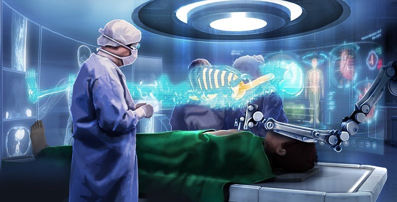
As surgeons are always on the lookout for ways to enhance their operating procedures, they are often among the first healthcare providers to adopt the latest technology.
Augmented reality (AR) is currently being used in clinical settings for medical training, intervention guidance and patient education.
Philips has developed image-guided therapies in partnership with Microsoft, to create a 3D holographic AR in healthcare environments for minimally-invasive procedures in future operating rooms.
It’s something Dr Morgan describes as a “promising approach”, and one which is receiving extensive investments due to the increase of smart connected devices integrating AR.
In its recently published study, Market Research Future reported the global healthcare AR market is expected to reach $1.32bn (£1bn) by 2023.
Dr Morgan said: “I see many applications for both training and real-time work.
“For example, the internal anatomy of the patient could be used as an overlay on the actual patient to help guide the surgeon.
“Interventional radiologists could use it to see a virtual ‘map’ of the patient’s vessels as they navigate the patient’s arteries during a procedure like treating an internal bleed or an aneurism [excessive swelling of the wall of an artery].”
Why Elsevier sponsored a neuroradiology symposium
Elsevier recently took part in the European Congress of Radiology in Vienna, Austria, during which it sponsored Dr Anne Osborn’s symposium, which was a huge success with over 30,000 attendees.
Dr Osborn is a professor of radiology at the University of Utah, and is recognised internationally for helping establish the field of neuroradiology, which deals with the head, neck, spine and the central and peripheral nervous system.
“In a course titled ‘An Excellence Masterclass,’Dr Osborn spoke on ‘Brain in Flame,’ discussing the role of aberrant brain inflammation in a broad spectrum of CNS pathology,” says Dr Morgan.
“This was followed by an interactive case-based session demonstrating the use of STATdx, Elsevier’s premier radiology decision support tool, to solve diagnostic dilemmas.
“Dr Osborn is a rock star in neuroradiology and wherever she goes a crowd always follows, making this a must-see event at the conference.”


