Solution uses deep learning technology allowing hospitals, pharmaceutical companies, and cancer centers to offer better and quicker diagnoses while developing more effective drugs and therapeutic interventions

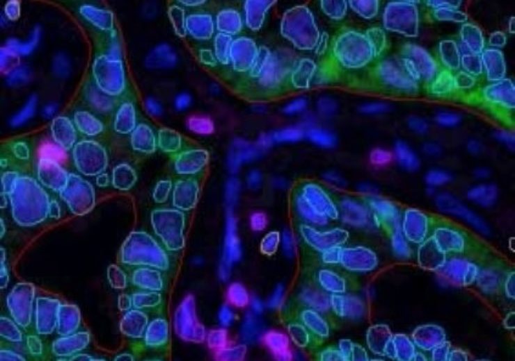
Image: RSIP Vision introduces image analysis solution. Photo: Courtesy of RSIP.
RSIP Vision, a global leader in artificial intelligence (AI), computer vision, and image processing technology, announced today that they are introducing an AI-based multiplex IF image analysis solution for precise results in tissue diagnosis. The new solution is based on a custom deep learning technology, allowing hospitals, pharmaceutical companies and cancer centers to better analysis and develop more effective drugs and therapeutic interventions.
Developing a precise AI-based approach to tissue analysis is critical to patient treatment since cellular behavior is largely impacted by the tissue microenvironment. The new solution allows precise analysis of marker expression allowing researchers to better understand the tissue landscape providing an exact indication of the immune status which could have a direct impact in therapy selection. This is driven by RSIP Vision superior ability to detect individual cells in tissue regardless of its morphology or staining intensity, combined with the ability to better separate merging cells.
“By utilizing AI and deep learning, we are able to provide physicians, hospitals and pharmaceutical companies a solution that has the highest proven accuracy for tissue analysis,” says Alan Jerusalmi, PhD, VP Business Development of Pharma Services at RSIP Vision. “High level precision will enable physicians to discover the best therapeutic strategy for patients in a much shorter period of time.”
The AI-based solution solves many of the challenges researchers and R&D teams face when analyzing multiplex images. It decreases false positives, significantly improves accuracy of nuclear detection and improves accuracy of phenotypic classification of cells.
“The contribution of the microenvironment to tumor progression has drawn considerable attention over the past years,” says Professor Ariel Munitz, Head of a leading innovative lab in the Department of Clinical Microbiology and Immunology at Tel Aviv University. “Using this new solution that provides the ability to visualize -in the tissue – multiple targets along with immune and cancer cell phenotyping, which is necessary to identify subsets of cells within the tumor microenvironment, providing major implications. Scientifically, it will provide new understandings on the complexity of the tumor microenvironment. Therapeutically, it will enable better stratification of patients and the development of tailored, combinatorial therapeutic intervention.”
Source: Company Press Release
