A non-invasive method to distinguish between skin cancer and benign lesions using smartphone and airport scanner tech has been developed by the Stevens Institute of Technology.
Researchers claim that using the new method could potentially cut the number of unnecessary biopsies by half.
According to the Centers for Disease Control and Prevention, skin cancer is the most common form of the disease in the US, and as the country’s older population grows, the number of new cases is expected to jump by as much as 2.1 million by 2030.
Director of the Stevens Bio-Electromagnetics Laboratory Negar Tavassolian said: “We’ve shown proof-of-concept that this technology can be used for rapidly detecting skin cancer.
“That’s a major step forward toward our ultimate goal of developing a handheld device, which would be safe to use directly on the skin for an almost instant diagnostic reading of specific kinds of skin cancer — including lethal melanomas — based on their individual reflectivity signatures.”
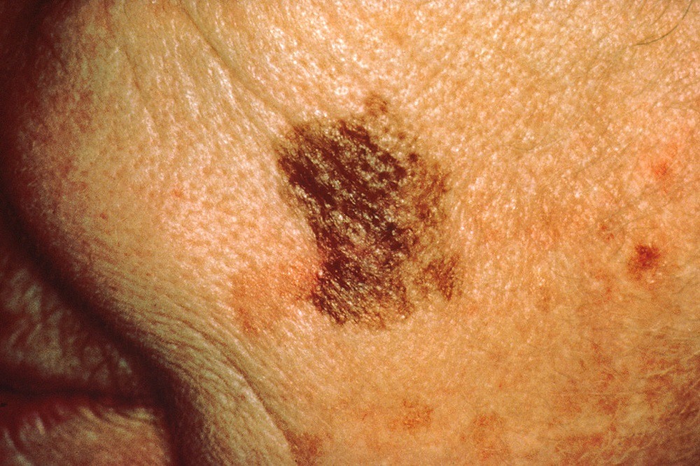
Melanoma is considered the most serious form of skin cancer. The disease often starts in the pigment-producing cells located in the deepest part of the outer layer of skin.
Changes begin to occur at this location long before people start seeing mole-like indications appearing.
How the new system uses millimeter wave rays to diagnose skin cancer
Non-invasive methods to detect skin tumours have already been produced, such as the device designed by a Rutgers University scientist that can quickly determine a skin lesion’s depth and potential malignancy, according to a report in Wiley Online Library.
Moreover, scientists from the University of British Columbia in Canada have developed a specialised microscope that has the potential to pinpoint the exact location of skin cancer as well as perform precise surgery without making incisions in the skin.
However, this new system uses millimeter wave radiation, which is longer than infrared waves or x-rays.
One of the handful of current uses of millimeter wave radiation is in airport scanners, and a variety of services on mobile and wireless networks, as it allows for higher data rates.
Millimeter wave rays penetrate certain materials and bounce off others, which is how airport security is notified of metallic objects such as keys carried by a person walking through a scanner.
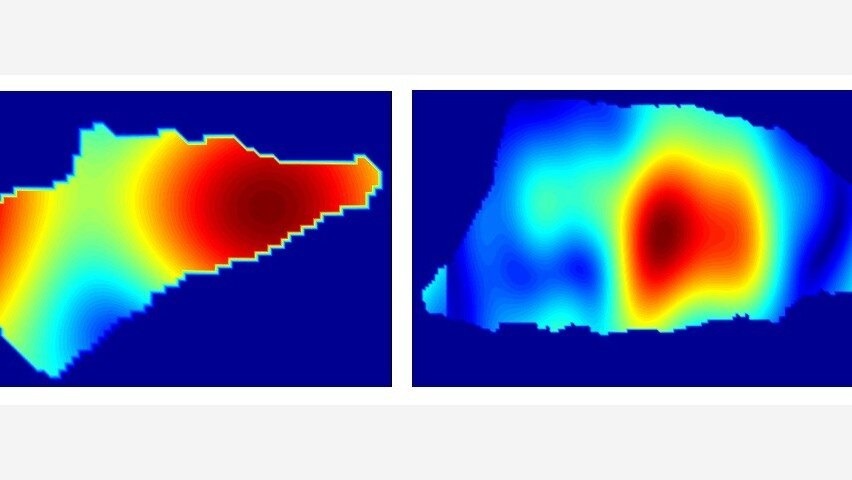
Therefore just as metal reflects more energy than the body — cancerous tumors reflect more calibrated energy than healthy skin — making it possible to identify diseased tissue by looking for “reflective hotspots.”
The scanners can then compose real-time 3D images of tumours that could help direct surgeons and eliminate the need for multiple trial-and-error biopsies to remove cancerous tissue.
In addition, the devices could be designed to automatically interpret images and present people with basic diagnostic information — triggering warnings if they need to get ssek medical advice.
“We could place these devices in pharmacies, so people can get checked out and go to a doctor for a follow-up if necessary,” said Tavassolian.
“People won’t need to wait weeks to get results, and that will save lives.”
Since it involves the same basic circuitry within a phone, it can therefore have an impact on the on the manufacturing costs making it quite inexpensive.
“It should be possible to keep manufacturing costs below $1,000, even at low production volumes.
“That’s about the same as the magnifying tools already used by dermatologists, and an order of magnitude cheaper than laser-based imaging tools, which also tend to be slower, bulkier and less accurate than millimeter-wave scanners.”
Titled High-Contrast, Low-Cost, 3-D Visualization of Skin Cancer Using Ultra-High-Resolution Millimeter-Wave Imaging, the study has been published in the September 2019 issue of IEEE Transactions on Medical Imaging.


