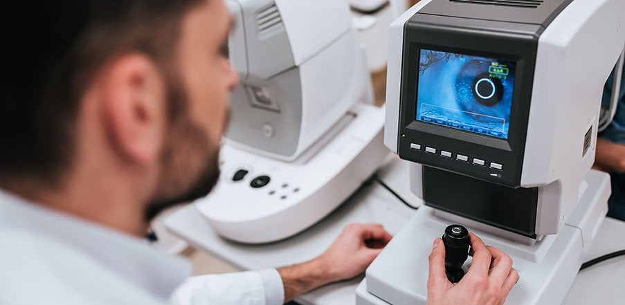
A team of scientists at Tohoku University, Japan has created a new deep learning (DL) model to enable automated screening of common eye disorders.
According to the report published in the journal Scientific Reports, the new lightweight DL is designed to identify disease-related features using images of eyes.
It can be trained with a small number of images, those with high levels of noise, and can be deployed on mobile devices, said the publication.
Tohoku University professor Nakazawa, associate professor Parmanand Sharma, Takahiro Ninomiya, and students from the University’s Department of Ophthalmology worked with professor Takayuki Okatani to develop the DL model.
Tohoku University Department of Ophthalmology professor and the study co-author Toru Nakazawa said: “There is always a trade-off between accuracy, speed and computational resources when it comes to DL models.
“Our developed model has better segmentation accuracy and enhanced model training reproducibility, even with fewer parameters – making it efficient and more lightweight when compared to other commercial softwares.”
According to the researchers, DL algorithms are predominantly task-specific and identify general objects such as humans, animals, or road signs.
The model requires accurate measurement of tumours, tissue volume, or other abnormalities, for self-monitory and tele-screening of diseases.
It would scan separate images and mark boundaries in a process called segmentation.
In addition, accurate prediction requires enhanced computational output, and rendering them is difficult to deploy on mobile devices.
The team obtained measurements of the foveal avascular zone, a region with the fovea centralis at the centre of the retina, for screening of glaucoma using low-resource devices.
In addition, the team aims to deploy a lightweight model for screening other common eye disorders and other diseases, in the near future.
Nakazawa added: “Our model is also capable of detecting/segmenting optic discs and haemorrhages in fundus images with high precision.”


