
Innovative technology provides accurate coronary artery 3D reconstruction from 2D angiography to be used for diagnosis, measurements, stent modelling, and characterization for procedural planning
Reconstruction of the coronary vasculature during angiography into a 3D model. (Credit: RSIP Vision.)
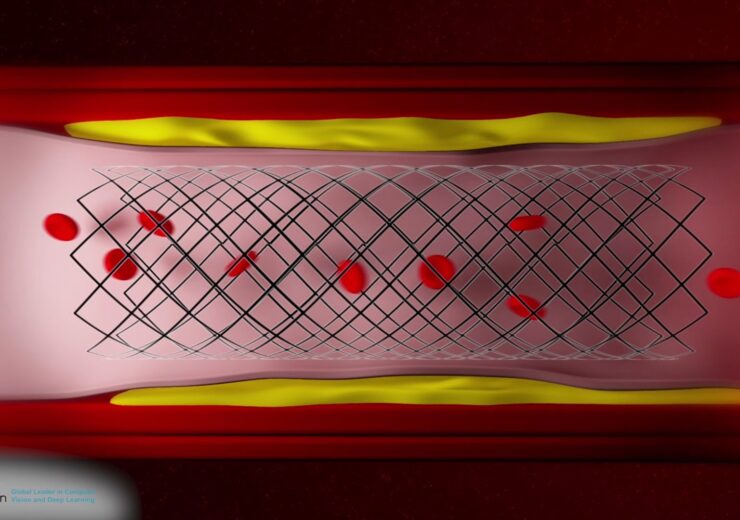
RSIP Vision, an experienced leader in driving innovation for medical imaging through advanced AI and computer vision solutions, today announces a new coronary artery modelling technology. This technology enables quick and accurate reconstruction of the coronary vasculature during angiography into a 3D model. This can be used for precise measurements of arterial length and diameter at any point, quantitative coronary angiography (QCA), including delineation of potential luminal obstructions, virtual stent positioning and vessel modification for intervention planning. This new vendor-neutral technology will be available to third-party C-Arm manufacturers and medical devices vendors, allowing an improved way to save lives and improve day-to-day clinical care.
“Coronary angiography is a common diagnostic medical procedure that may involve a therapeutic/interventional stage if significant pathological findings are encountered,” said Ron Soferman, CEO at RSIP Vision. “Following the diagnostic imaging step, the data can be reconstructed to a 3D model and used for detection, diagnosis, and characterization of coronary artery stenosis. This technology will be useful in percutaneous coronary intervention (PCI) planning, reducing the need for additional interventions and decreasing the chance of adverse events due to incorrect stent selection.”
Developing capabilities for additional interventions during PCI planning requires utilization of state-of-the-art deep learning (DL) algorithms combined with classic computer vision methods to create a 3D model of the coronary arteries accurately, rapidly, and automatically. RSIP Vision’s model can be used to better visualize artery structure and measure vessel dimensions in points-of-interest. More advanced capabilities, enabled using DL, are stenosis detection, 3D QCA, and can even be the baseline for computerized FFR measurement. Additional modules can show anticipated post-procedural arterial modification/outcome because of stent placement or place a virtual stent in the desired position within the coronary artery. These uses may supplement already existing clinical and imaging tools and, in some cases, even replace invasive cardiac measurements, and give the physician a better pre-procedure planning tool for stent selection.
“Pre-procedural PCI planning and intra-procedural analysis, appraisal and proper execution form the pillars of an uneventful invasive coronary intervention,” said Dr. David Yakobi, Board Certified Cardiac Surgeon/Medical Consultant. “Automated technologies that would allow better anatomical and pathological visualization and appreciation, as well as AI-based algorithms for coronary lesion characterization, will augment the procedural success, decrease the complication rate and (based on a very broad scientific literature) have the potential of increasing stent patency rate and patients’ long-term survival.
“This module from RSIP Vision has the potential of becoming an automated, human error and bias free tool, for classifying Coronary Artery Disease based on the SYNTAX score and basically behaving as an independent ‘Heart Team’ for decision-making purposes between PCI and CABG (Coronary Artery Bypass Graft) surgery.”
Source: Company Press Release