Dutch medical technology firm Royal Philips has secured the US Food and Drug Administration 510(k) clearance for its Philips SmartCT application software.
SmartCT is an image acquisition, visualisation and measurement software, part of the Philips Image Guided Therapy System Azurion, which was launched in September last year.
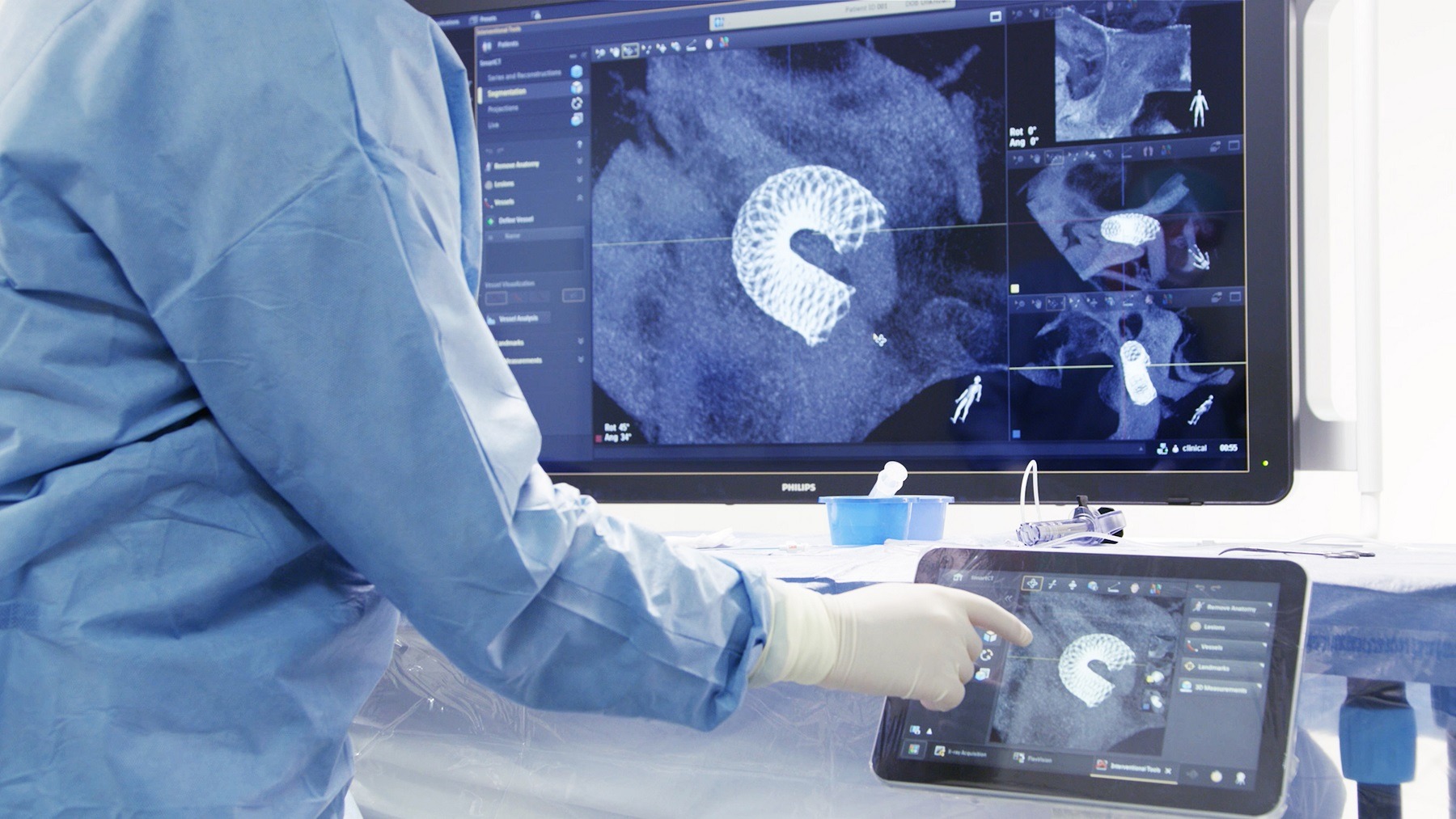
The software provides interventionalists with CT-like 3D images, dubbed Cone Beam CT, to support diagnosis, therapy planning, treatment and follow-up for interventional radiology procedures.
Philips image guided therapy systems general manager Ronald Tabaksblat said: “A key part of our image-guided therapy strategy is to combine high-quality, low X-ray dose imaging with a superior user experience that allows interventional radiologists to diagnose and treat patients as part of smoother, safer and less interrupted workflows.
“Philips SmartCT is a major step forward in 3D imaging, enhancing confidence in the interventional suite and supporting key elements of the quadruple aim of better patient outcomes, enhanced patient and staff experiences and lower cost of care.”
The company said that its Azurion image-guided therapy platform integrates important lab systems and tools required for complex interventional procedures into a laboratory environment.
SmartCT brings intuitive touchscreen control of the Azurion platform to the table-side, within the sterile area of the interventional lab.
The solution eliminates the need for clinicians to leave the sterile field and step into a control room, along with supporting rapid and better-informed decision making.
SmartCT can be used for angiography, neurology, soft-tissue imaging, and guidewire or catheter navigation, and supports the treatment of aneurysms, vascular diseases and liver tumours.
Philips said that its offering guides users through the image acquisition process and enables them to review and interact with the acquired CT-like 3D images using 3D visualisation.
Also, it features intuitive two-point distance measurements on 3D images, and the capability to select and store optimum projection angles for recall during procedures.
Hôpitaux Universitaires Henri-Mondor radiology and interventional radiology chief Hicham Kobeiter said: “Changing to a new technology can be challenging, but if the system itself can show you the way, it makes it much easier to adopt new advances.
“SmartCT leads you through each step of the procedure, bringing us more confidence and more precision across cardiovascular, oncology and emergency cases.”


