KARL STORZ Endoscopy, a US-based endoscopy, imaging, and operating room integration solutions provider, has announced the results from a prospective, randomized multicentre trial to evaluate use of near-infrared fluorescence cholangiography (NIFC) to detect extrahepatic biliary structures.
The trial was conducted at seven centres in the US, Germany, Italy, Argentina, and Japan, involving four continents, with an aim to evaluate the capability of NIFC in increasing the detection rate of extrahepatic biliary structures relative to white light.
One of the study authors, Raul Rosenthal said: “We still experience complications when performing laparoscopic cholecystectomy. Since the introduction of laparoscopy in 1989, the incidence of common bile duct injuries has doubled from 0.4% to close to 0.8% of those undergoing this approach.
“Bile duct injuries, despite being rare, can have life‐threatening connotations, and in 97% of the cases they are due to a visual misperception: the surgeon believes that he or she is working on the cystic duct, but in reality is working on the common bile duct.”
Use of NIR/ICG imaging technology with ICG offers enhanced results
According to the KARL, laparoscopic cholecystectomy is a common procedure, where surgeons face problems in identifying the biliary tract anatomy, and accurate identification and prevention of potential injury to the cystic duct and common bile duct are primary challenges to surgeons.
The study marks the first randomised trial to compare laparoscopic cholecystectomies performed under white light, with those performed under combined white light and near-infrared (NIR), after injection with indocyanine green (ICG) dye.
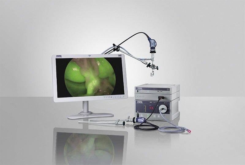
In addition, the procedures were performed using the NIR/ICG Imaging System, an innovative extension of the IMAGE1 S System, a single adaptable video architecture featuring modular CCU.
The system facilitates surgeons to perform minimally invasive surgery using conventional endoscopic white light or using NIR/ICG fluorescence imaging for enhanced visual assessment of vessels, blood flow, and related tissue perfusion.
KARL STORZ said that the use of NIR/ICG imaging technology in combination with ICG offers enhanced visualisation, evaluation of organs, associated tissues, bile duct, and also help assessing perfusion and associated defects.
Rosenthal added: “The results are staggering, because not only were the surgeons that utilized both white light and near-infrared light able to visualize the biliary tract anatomy two to three times better than those who utilized white light alone, but also the group that utilized only white light had two common bile duct injuries in 300 cases, while the group that used both white and the near-infrared with fluorescent imaging light had no injuries.”


