This technology provides an x-z plane scan using a light-sheet for a whole 96/384/1536-well microplate, allowing users to obtain 3D fluorescence images of whole wells in xy: 2-3μm and z: 6-7μm voxel resolution for ≤ 300μm thickness from the well bottom within a few minutes per colour.
This technology also allows for ultra-high level separation of cell fluorescence signals from the background, which makes it possible to obtain fluorescence cell images in the cell culture mediums containing serum and with fluorescent dyes (i.e., no need to wash out fluorescent dyes).
In drug discovery and development, there is a rapidly growing need to use more physiologically-relevant in vitro cell culture models. These include primary cells, patient-derived cells, human iPSC-derived disease-modeling cells, co-cultures of different types of cells, and 3D cultures such as spheroids and organoids.
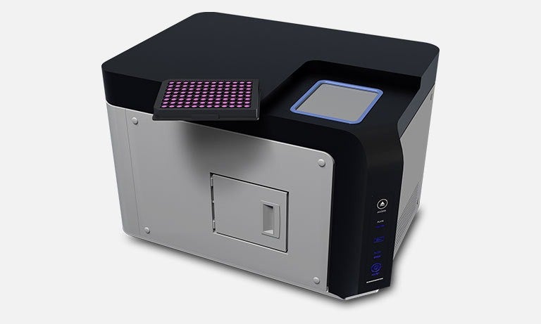
To develop and advance phenotypic assays and screenings using these physiologically-relevant cellular models, new types of instruments needed to be developed to facilitate high-throughput fluorescence imaging and measurement of heterogeneous 2D and 3D cell cultures. To meet this need, we have developed ZyncscanTM technology that performs an x-z plane scan utilizing a light-sheet for a microplate.
High-speed fluorescence cytometry for 2D monolayer heterogeneous cell culture, A single 120-second scan (one color) for a whole 96/384/1536-well microplate enables fluorescence cytometry of all individual cells in all wells.
Single spheroid analyses using depth information (diameter of spheroid; ≤ 200 μm), A single scan (in just a few minutes) for a microplate enables fluorescence images of spheroids (single or multiple spheroids in a well) in all wells in a 96/384/1536-well microplate. From the images, the entire fluorescence intensity, thickness and volume of each individual spheroid are estimated.
Fluorescence images in 3D view (≤ 300 μm), With a single scan (a few minutes) obtain 3D fluorescence images (300 μm from the bottom of a well) of all wells in a plate. The optical resolution of the image is comparable with the 2x objective of a conventional fluorescence microscope in the x- and y-axes and a 10x objective of confocal fluorescence microscope in the z-axis.
In-medium / No dye washout measurement, Measure cellular fluorescence in the medium containing serum and with fluorescent dyes (no need to wash out fluorescent dyes), allowing cells to remain healthy throughout the experiment.
Source: Company Press Release.


