
The researchers found that use of PVi, as a dynamic, continuous method of managing fluid administration, led to patients receiving less fluid and having fewer B-lines (a measure of EVLW), with no significant decrease in postoperative perfusion or oxygenation, compared to use of a central venous pressure (CVP)-guided fluid administration protocol
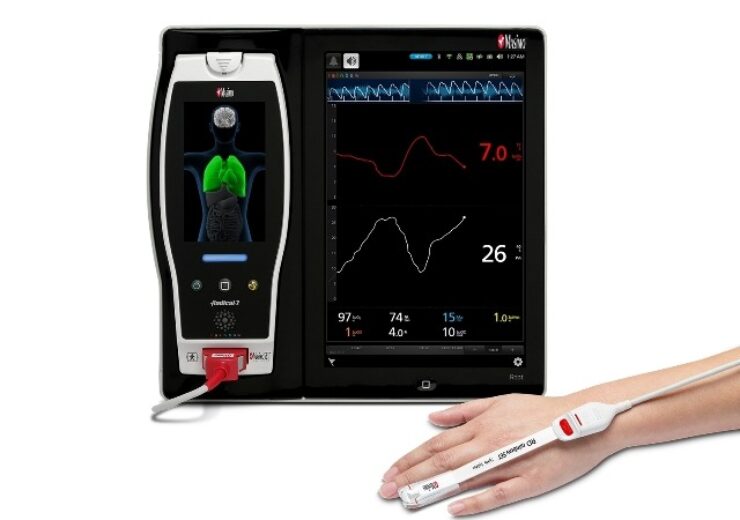
Masimo Root with PVi and SpHb. (Credit: Business Wire)
Masimo (NASDAQ: MASI) today announced the findings of an abstract presented at ANESTHESIOLOGY 2021 in San Diego, in which Drs. Anita Kulkarni and Arti Dalal at the Rajiv Gandhi Cancer Institute in New Delhi, India evaluated the use of noninvasive, continuous Masimo PVi® as part of goal-directed fluid therapy (GDFT) to guide intraoperative fluid administration during major oncosurgery in which lung ultrasonography (LUS) is used to diagnose extravascular lung water (EVLW).
The researchers found that use of PVi, as a dynamic, continuous method of managing fluid administration, led to patients receiving less fluid and having fewer B-lines (a measure of EVLW), with no significant decrease in postoperative perfusion or oxygenation, compared to use of a central venous pressure (CVP)-guided fluid administration protocol.
Noting that conventional invasive methods of guiding fluid administration (such as CVP) during major oncosurgery may increase EVLW, which can lead to postoperative cardiorespiratory complications, the authors investigated whether use of a noninvasive, dynamic, continuous method—GDFT guided by Masimo PVi—could improve fluid management by reducing the amount of fluids administered and B-lines as measured by LUS. To evaluate PVi, they compared two groups of adult patients undergoing major oncosurgery with LUS: a group of 60 patients whose fluid administration was guided by PVi as part of GDFT and a group of 59 patients whose fluid administration was guided by CVP. Their primary outcome was detection of EVLW indicated by the total number of B-lines, and their secondary outcome was adequacy of perfusion by lactate levels at the end of surgery. The authors also measured arterial blood gas samples at baseline and the end of surgery to evaluate oxygenation.
Comparing the amount of fluids administered, the researchers found there were significantly fewer total crystalloids given in the PVi group (1875.8 ± 593.9 ml) compared to the CVP group (2132.6 ± 504.5 ml; p = 0.012), as well as significantly less colloid provided in the bolus (PVi group: 584 ± 358.5 ml; CVP group: 778.2 ± 242.2 ml; p = 0.001). They found that 23.3% of PVi-group patients had “mild” B-lines and 5.0% “moderate” B-lines, compared to 44.1% mild and 5.1% moderate in CVP-group patients (p = 0.042). At the end of surgery, they found that in the CVP group, there was a statistically significant decrease (p < 0.05) in partial pressure of oxygen in arterial blood (PaO2) and the ratio of arterial blood oxygen partial pressure to fractional inspired oxygen (PaO2/FiO2), compared to baseline levels; in the PVi group, the parameters were comparable to baseline after surgery, with no significant difference.
The authors concluded, “We recommend lung ultrasonography after completion of major oncosurgeries to detect EVLW and intraoperative PVi-guided GDFT as these patients received less fluids, had less B-lines (mild) and no decrease in PaO2 or PaO2/FiO2 levels at [the] end of surgery.”
In the U.S., PVi is cleared as a noninvasive, dynamic indicator of fluid responsiveness in select populations of mechanically ventilated adult patients.
Source: Company Press Release